Kiemelkedő biztonság CBCT felvétel
3 Dimenziós Cone Beam CT a fogbeültetés és csontpótlás szolgálatában
A 3 dimenziós képalkotás a fogbeültetési és csontpótlási műtéti eljárások alapvető diagnosztikai eszköze. A fogbeültetés és a csontpótlás megtervezése akkor lesz sikeres és megbízható, ha az orvos a csontot minden irányból megfelelően feltérképezheti, lemérheti. A mérések alapján a szájsebész pontosan meghatározhatja a beültetendő fogimplantátum méretét, elkerülheti az idegeket és fontos anatómiai üregeket, így a fogpótlás vagy csontbeültetés műtéte lényegesen biztonságosabb.
Fontos Önnek a maximális biztonság a fogbeültetésben és a csontpótlásban?
Mert nekünk igen! Mi szeretnénk tökéletesen biztosak lenni abban, hogy az Ön fogbeültetése és csontpótlása kapcsán is a legnagyobb biztonsággal, körültekintéssel és szakértelemmel jártunk el! Ezért alkalmazzuk a kiemelkedő képminőséget biztosító 3 dimenziós CT készüléket a műtéti tervezésben és a kivitelezés során is. Fogbeültetésben gondolkodik? Kérjen időpontot konzultációra!
Miért jó Önnek? Mi az előnye a CBCT-nek a hagyományos röntgennel szemben?
A CBCT a Cone Beam Computer Tomográf rövidítése, mely kifejezetten az arc, állcsont régióra kifejlesztett, speciális 3 dimenziós röntgenkép készítését jelenti. Míg a panoráma röntgen csak 2 dimenzióban mutatja meg a csontot, addig a 3 dimenzió teljes térbeli leképezést jelent. A 3. dimenzió, azaz a mélység megjelenítése hatalmas többlet információhoz juttatja a diagnosztizáló szakorvost. Olyan csontrészletek is megjeleníthetőek, melyek a 2 dimenziós, azaz hagyományos röntgenfelvételeken egyáltalán nem vagy elmosódottan látszódtak. A 3 dimenziós CT segítségével tehát az orvos virtuális valóságában, minden kiterjedésében vizsgálhatja az Ön csontjának formáját, minőségi mutatóit. A képen a csontokat virtuálisan elforgathatja, lemérheti, tanulmányozhatja a belső felszíneket, így korrektebb és biztosabb diagnózis és műtéti kivitelezés valósulhat meg. A 3 dimenziós CT felvétel segítségével kivitelezett fogbeültetés és csontpótlás ezért nagyobb biztonságot jelent az orvos és a páciens számára egyaránt.
“Itt nem én vagyok a kísérleti nyúl!”
“A fogbeültetés sikeraránya 98%. A lényeg a megfelelő tervezésen és előkészítés. Nem lehet bárhova és bármilyen céllal implantálni, pontosan meg kell tervezni a beültetett implantátumra kerülő fogpótlást is már az elején. A fogpótlás megtervezése után következik maga a műtéti tervezés. Ennek során 3 dimenziós CT felvételt alkalmazunk, hogy megfelelő képünk legyen a csont belsejéről…”
Valóban igen alacsony sugárdózissal jár a CBCT készítése?
Sok páciensünk érdeklődik az őt érő sugárdózis mennyisége felől. Sokan a hagyományos, nem fogászati alkalmazású, úgynevezett orvosi CT felvételek paramétereit feltételezik, amelyek készítése során valóban jelentős dózis érhette a pácienst. A Cone Beam CT technológiája azonban lényegesen eltér a hagyományos orvosi CT technológiától. Az eltérő technológiának köszönhetően, a Cone Beam CT használatakor a nem fogorvosi felhasználású CT felvételek során kibocsátott sugárdózisnak körülbelül 1%-a éri a pácienst.
Ez az egy százaléknyi dózis nagyságrendileg megegyezik a fogászatban eddig használt, hagyományos 2 dimenziós röntgen eljárások sugárterhelésével. További jelentős különbség, hogy a szkennelés a hagyományos orvosi CT-vel perceket vesz igénybe, míg CBCT-vel csupán 20-40 másodpercet.
Miért lényeges a 3 D CBCT a fogbeültetés és csontpótlás során?
A háromdimenziós nézetek megkönnyítik a fogimplantátumot befogadó csont feltérképezését. A csontviszonyok valós ismeretében pontosan megtervezhető a megfelelő implantátum, vagy implantátumok elhelyezkedése. A pontos tervezés kisebb kockázatot, nagyobb biztonságot jelent a műtéti kivitelezés során. A szájsebész már előre “ismeri” a csontot, még mielőtt a műtét elkezdődne. Biztos abban, hogy mit talál a fogíny alatt, tudja mire számíthat, jobban felkészülhet a teljes folyamatra. A műtét után lehetőség van azonnali kontrollálásra.
Miért lényeges a 3 D CBCT a fogágybetegségek kezelése során?
A többirányú felvételeknek köszönhetően lehetővé válik a fogínytasak dimenziók pontos feltérképezése. A pontos felmérés kiemelkedően fontos a fogpótlások megtervezésénél. Az orvos sokkal biztosabb képet kap arról, hogy egy adott fog megtartható e és biztonsággal építhető e rá a megtervezett fogpótlás. A biztos alapokon nyugvó tervezés hosszabb távon stabilan funkcionáló fogpótlást jelent.
Miért lényeges a 3 D CBCT a gyökérkezelés során?
A Cone Beam CT segítségével pontosan megállapítható a gyökércsatornák lefutása, a gyökércsúcson kialakult gyulladás jobban feltérképezhető. Biztosabban megállapítható, hogy egy adott fogat érdemes e egyáltalán elkezdeni gyökérkezeléssel megmenteni, vagy inkább a foghúzás felé érdemes lépni. A CT felvétel segítségével a foggyökerek biztonságosabban kikezelhetőek, több fogat menthetünk meg.
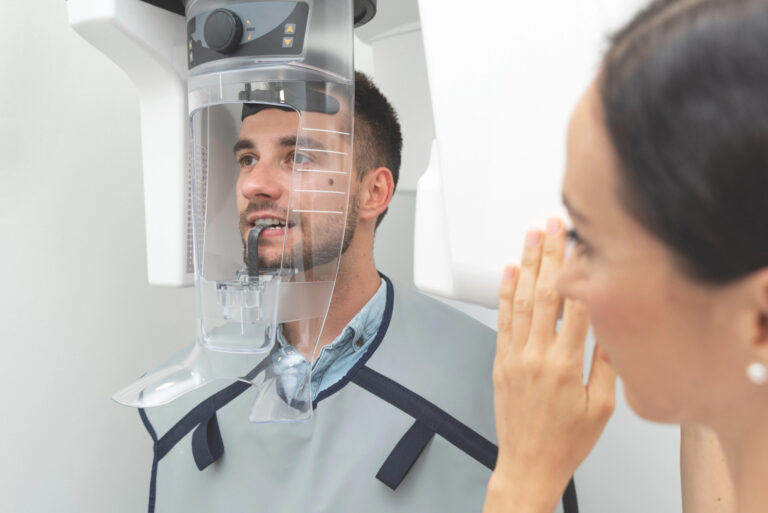
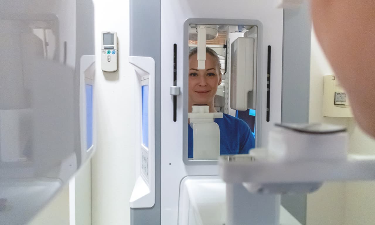
Hogyan készül a CBCT felvétel?
A CBCT felvétel álló helyzetben készül. A fej körül forog el a készülék, a felvétel ideje alatt nem szabad mozogni. A felvétel teljesen fájdalommentes. A testet ólomkötény védi a sugárzástól.